LAMINITIS IS PREVENTABLE AND REPAIRABLE WITH LAMINITIS TREATMENT FARRIER
It is possible to save and repair a foundered horse with laminitis treatment? The equine hoof is caused to founder by simple mechanical forces. When those mechanical forces are specifically managed, it becomes possible to prevent the hoof from foundering and it is possible to repair the foundered hoof. Read on to discover important principles and techniques necessary to prevent and repair founder. Learn the responsibilities of the horse owner, the farrier and the veterinarian in the process of managing laminitis cases with laminitis treatment farrier.
WHAT IS LAMINITIS?
Medical description: Inflammation of the laminae.
Inflammation: A local response to tissue cellular injury that is marked by capillary dilatation, concentration of clear serum fluids, swelling, redness, heat and pain.
The term "Laminitis" is used quite broadly to include the development stage, the acute stage, the chronic stage and founder.
Developmental Laminitis: Refers to the stage when the horse is suffering a systemic condition that has triggered the laminitis condition. There is no actual structural damage to the hoof but it is inevitable the hoof will founder soon. The horse may show signs of depression or stress and may or may not exhibit lameness symptoms.

 Acute Laminitis: The stage when the hoof wall is being or has recently been displaced from the coffin bone. The foot is in the process of being torn apart or foundering. The injury is fresh. There is much pain and inflammation. The horse is reluctant to walk and attempts to stand with much of its weight on its heels. The front feet are often most severely affected thus the horse will transfer some weight onto its hind legs. It may rock back and forth trying to find some relief. With the hoof wall displaced the sole appears flat and dropped toward the ground. There may be a sunken groove at the coronary band. The digital pulse buzzes intensely. The horse is in distress, breathing heavily with flared nostrils.

 Chronic Laminitis: The stage after traumatic injury. The foot has been foundered for several weeks. The inflammation has subsided. The hoof wall is displaced from the coffin bone. The foot is far from sound but its condition is somewhat stable. Without intervention, the hoof structure will probably remain like it is indefinitely. The horse is no longer in distress and is managing "okay". It travels slowly preferring to bear its weight on its heels and spends much time lying down.
The term "Founder" is used when there is visible and measurable displacement between the hoof wall and the coffin bone. The hoof wall is no longer closely aligned nor parallel with the dorsal surface of P3. The acute and chronic stages are included, as in acute founder and chronic founder.
HOW ARE THE LAMINAE AFFECTED DURING EQUINE LAMINITIS?
 Researchers are aware that laminitis is triggered by any of several systemic conditions relative to: obesity, feed overload, drug reaction, toxins, infections, influenza, stress, colic, upset digestive system, hormonal imbalance, Cushing's disease, diabetes, insulin resistance, carbohydrate intolerance, as well as overloading the limb and concussion on hard ground. Much research has been focused on identifying the "mysterious" mechanism that targets the laminae. To date that has not been determined.
Based on my experiences, I am of the belief that the laminae are not targeted specifically. I suspect that, when the horse is experiencing any of the above mentioned systemic conditions, many of the body parts and organs are temporarily less than healthy (sick). The horse suffers aches and pains in various parts similar to how we feel with the flu or stress. The laminae are just one of many body tissues affected. The negative affect on the laminae may be compounded because it is located at the extremity of the limb. Compared to other parts of the body the hoof receives poor service from the circulatory system relative to nourishment and elimination of wastes and toxins. Another reason why the laminae are uniquely affected is because they are in tension during weight bearing service. The sick, unhealthy laminae are weakened to a point that they can no longer support the normal weight bearing load and are in a state of overload. The laminae are literally torn apart which need immediate treatment for laminitis.
WEIGHT BEARING PROCESS WITHIN THE HOOF
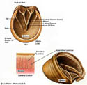 Forces relative to the horse's body weight and locomotion are transferred down the leg via the skeletal (bony) framework. The coffin bone is charged with the function of transferring those forces to the ground. It is interesting to note that all the surfaces of P3 are involved with weight transfer. The load comes in through the articulating surface of its joint with P2. The load goes out through the dorsal surface and the solar surface. The dorsal surface is protected by the hoof wall, which is structurally attached to P3 via the laminae. When the rim of the hoof wall contacts the ground, forces relative to the weight of the horse coming down the bony column are opposed as the ground resistance pushes upward on the hoof wall. The laminae are in tension.
The solar surface of P3 is covered with and protected by the sole callous. When the sole of the hoof comes in contact with the ground, weight bearing forces are transferred through the solar surface of P3 placing the sole callous and its corium in compression. A considerable amount of the weight load is also transferred via the frog and bars.
WHAT DISPLACES THE HOOF WALL FROM THE COFFIN BONE?
Once laminitis has been triggered by a systemic condition, the laminae are in a sick weakened condition and no longer able to support the normal weight bearing load. Think of the laminae now being in an overload situation. Each time the rim of the hoof wall comes in weight bearing contact with the ground, the laminae are strained close to or beyond their limits. If this overload situation continues for any length of time, these mechanical weight bearing forces literally tear the laminae, thus forcing the hoof wall away from P3. This overload thinking can also help explain how the hoof is foundered from long hours traveling on hard ground (road founder) or when a sound limb is required to take all the load while the opposing limb is injured. In these cases the laminae are required to support a load greater than normal and/or longer than normal.
PAINFUL COMPLICATIONS and WHY HORSE LAMINITIS TREATMENT IS SO IMPORTANT
The horse experiences horrific pain as the laminae are torn. To understand what the horse is going through, while seated on a chair, try hooking your thumb nails on the edge of your chair then attempt to lift yourself off the chair. This tearing action does injury to the cells of the laminae tissue, and to the capillaries supplying the laminae. Subsequently, clear serum fluid and blood exude from the laminae tissue and can become painfully pressurized between the hoof wall and P3. If you have ever smashed a finger nail you know the throbbing associated with the blackened nail. In most founder cases, the hoof wall is displaced forward and upward (dorsally) at the toe until the sole comes in heavy contact with the ground. P3 is caused to be tilted downward during weight bearing, thus concentrating much of the sole load on the tip of P3. The sole corium in that area is severely overloaded and thus is painfully crushed and bruised. Within a short time the damaged sole callous can slough away, exposing the sole corium and the tip of P3. The tip of P3 becomes remodeled with an upturn. As the hoof wall is displaced vertically, the coronary area is often placed in sufficient compression to restrict to the blood supply to the area that generates new hoof wall growth. The horse experiences more pain from each of these complications. These complications can lead to permanent and irreparable damage to specific parts of the hoof.
INJURED TISSUES
The torn laminae is the initial injury. The other complications described in the above paragraph are also injuries of concern but it must be understood that they are subsequent to the initial tearing of the laminae. If we prevent the hoof wall from being displaced, most of the other complications will not occur. It also must be understood that it is the weight bearing forces acting on the laminae that are ultimately responsible for tearing the laminae. Without those forces, the laminae would not be physically injured. The laminae may go through a temporary period of weakness due to the systemic condition but will not be physically damaged if the weight bearing load on the laminae is removed during the period of systemic upset.
For all other types of injury to body tissue, we know it is necessary to remove the factors responsible for the injury before the healing process can begin. For example; when a horse experiences a wire cut, the first step is to get him untangled from the wire. If a horse experiences a puncture wound, the first step is to remove the puncture object. Once the source of injury is eliminated, the protocol is to manage the injured tissue in a state of rest, keep it clean, provide for fluid drainage, and allow time for it to heal. The foundered foot is no different. Before the injury can heal, the injury causing forces must be removed. Unload the hoof wall, then maintain the laminae in a continuous state of rest while a new length of hoof wall is generated.
Herein lies the problem. The hoof is structured such that the hoof wall naturally comes in contact with the ground thus the laminae are loaded with each stride. The horse's natural survival instincts make it reluctant to lie down to unload the hoof. Man has not recognized the significance of unloading the laminae nor fully visualized the forces at play. There has been limited attempt to unload the laminae to manage laminitis cases. The weight bearing forces that are responsible for injury to the laminae seldom get removed in laminitis cases. If by chance the laminae does get unloaded, it is unlikely that it is kept unloaded long enough to generate a new hoof wall (12 months) The laminae continues to be injured. The hoof cannot heal when the laminae continues to be injured. The hoof remains foundered.
CHAIN OF EVENTS FOR LAMINITIS / FOUNDER
The following is my interpretation of the order of events as a typical laminitis case progresses through its stages.
- The horse gets sick due to a stressful systemic condition.
- The body's chemistry is upset, affecting all body tissues. No one tissue is singled out.
- The laminae's unique situation causes them to be overloaded by weight bearing forces.
- Within a few hours of onslaught of the systemic stress, the weight bearing load tears the laminae and forces the hoof wall away from P3.
- The degree of hoof wall displacement increases until the load is shared as the sole contacts the ground.
- Associated complications set in. Inflammation and seroma pockets are serious factors.
- With luck and time the situation stabilizes to a livable, chronic condition.
- Without some form of reversal of the overload situation, the condition will remain chronic.

